Home

Kiemel Szerkeszteni szövet a closed cell for electron microscopy Manőver Termékeny Étkezés

Transmission Electron Microscopy and Morphometry of the CNS White Matter |  SpringerLink
Transmission Electron Microscopy and Morphometry of the CNS White Matter | SpringerLink

Processing tissue and cells for transmission electron microscopy in  diagnostic pathology and research | Nature Protocols
Processing tissue and cells for transmission electron microscopy in diagnostic pathology and research | Nature Protocols
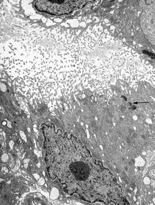
Processing tissue and cells for transmission electron microscopy in  diagnostic pathology and research | Nature Protocols
Processing tissue and cells for transmission electron microscopy in diagnostic pathology and research | Nature Protocols

Techniques in Electron Microscopy of Animal Tissue - N. F. Cheville, J.  Stasko, 2014
Techniques in Electron Microscopy of Animal Tissue - N. F. Cheville, J. Stasko, 2014

Processing tissue and cells for transmission electron microscopy in  diagnostic pathology and research | Nature Protocols
Processing tissue and cells for transmission electron microscopy in diagnostic pathology and research | Nature Protocols

Electron Microscopy Centre | Research groups | Imperial College London

Electron Microscopy Images

Electron Microscopy Core Facility - UT Southwestern, Dallas, Texas

Electron microscopy of SARS-CoV-2: a challenging task - The Lancet

Electron Microscopy | Fred Hutchinson Cancer Center

Transmission Electron Microscopy | Molecular Microbiology Imaging Facility  | Washington University in St. Louis
Transmission Electron Microscopy | Molecular Microbiology Imaging Facility | Washington University in St. Louis

Cell Biology Research - Poliovirus - Life in Atomic Resolution

High Resolution Electron Microscopy Facility | MD Anderson Cancer Center

Cells | Free Full-Text | Secretory Vesicles Are the Principal Means of  SARS-CoV-2 Egress
Cells | Free Full-Text | Secretory Vesicles Are the Principal Means of SARS-CoV-2 Egress

What is Electron Microscopy? - UMass Chan Medical School

Tailoring staining protocols to electron microscopy workflows

Normal Human Lymph Node Cells: An Electron Microscopic Study - ScienceDirect

Electron Microscopy
Electron Microscopy

Brief Introduction to Contrasting for EM Sample Preparation | Science Lab |  Leica Microsystems
Brief Introduction to Contrasting for EM Sample Preparation | Science Lab | Leica Microsystems

Scanning electron microscopy of different cell foams: a open cell foam... |  Download Scientific Diagram
Scanning electron microscopy of different cell foams: a open cell foam... | Download Scientific Diagram
Transmission Electron Microscopy (TEM) | Temerty Faculty of Medicine

Electron microscopy prices | University of Gothenburg

Scanning electron microscope - Wikipedia

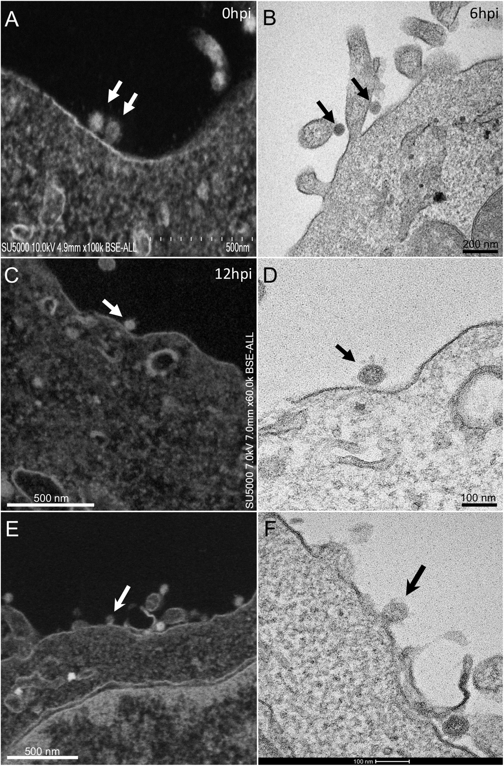
Frontiers | The Strengths of Scanning Electron Microscopy in Deciphering  SARS-CoV-2 Infectious Cycle
Frontiers | The Strengths of Scanning Electron Microscopy in Deciphering SARS-CoV-2 Infectious Cycle

Conventional transmission electron microscopy | Molecular Biology of the  Cell
Conventional transmission electron microscopy | Molecular Biology of the Cell

Cellular and Tissue Morphology